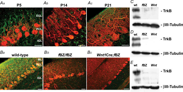
Figure 1. TrkB expression during cerebellar development under normal, TrkB-hypomorphic and TrkB-null conditions.
A, calbindin (red) and TrkB (green) immunostaining in the cerebellar cortex at postnatal day (P)5 (a), P14 (b), and P21 (c) in wild-type mice. Scale bars (Aa and b), 10 μm; (Ac), 20 μm. B, calbindin (red) and TrkB (green) immunostaining of wild-type (a), fBZ/fBZ (b), and Wnt1Cre;fBZ/fBZ (c) cerebella. Scale bars, 20 μm. C–E, Western blots of homogenized cerebella from wild-type (wt), fBZ/fBZ (fBZ), and Wnt1Cre;fBZ/fBZ (Wnt) mice at P4 (C), P14 (D), and P21 (E). TrkB expression was normalized to βIII-tubulin.

