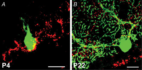
Figure 6. VGluT2 immunostaining reveals CF terminals and their translocation during cerebellar development.
A, innervation field of climbing fibres on a multiply innervated, Lucifer-yellow-filled Purkinje cell (green) in a P4 wild-type cerebellar slice. Climbing fibre terminals are labelled with a VGluT2 antibody (red) and restricted to the Purkinje cell soma (colocalization: yellow). Scale bar, 20 μm. B, by P22, the climbing fibre innervation field has translocated to the dendrite of this mono-innervated Purkinje cell. Scale bar, 20 μm.

