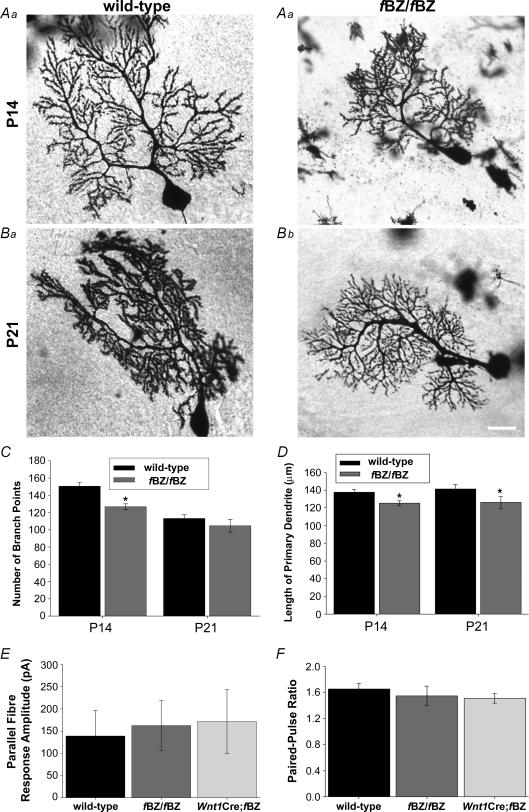
Figure 9. Parallel fibre synaptic transmission is normal at P20–24 in TrkB mutant mice despite an earlier delay in Purkinje cell dendritic development.
A and B, examples of Golgi-impregnated Purkinje cells from wild-type (a) and fBZ/fBZ (b) at P14 (A) and P21 (B). Scale bar, 20 μm. C, total number of dendritic branch points. There are significantly fewer branches of fBZ/fBZ mice at P14 (*P < 0.001 by one-way ANOVA), but not at P21 (P= 0.079 by one-way ANOVA). D, the length of the primary dendrite is shorter in fBZ/fBZ mice than wild-type mice at both P14 and P21 (P14 and P21: *P < 0.001 by one-way ANOVA). E, average maximum amplitude of parallel fibre responses from wild-type, fBZ/fBZ and Wnt1Cre;fBZ/fBZ mice was comparable (P= 0.930 by one-way ANOVA). F, the ratio of parallel fibre-activated paired-pulse facilitation in Purkinje cells from wild-type, fBZ/fBZ and Wnt1Cre;fBZ/fBZ cerebellum was also comparable at P20–24 (P= 0.632 by one-way ANOVA).

