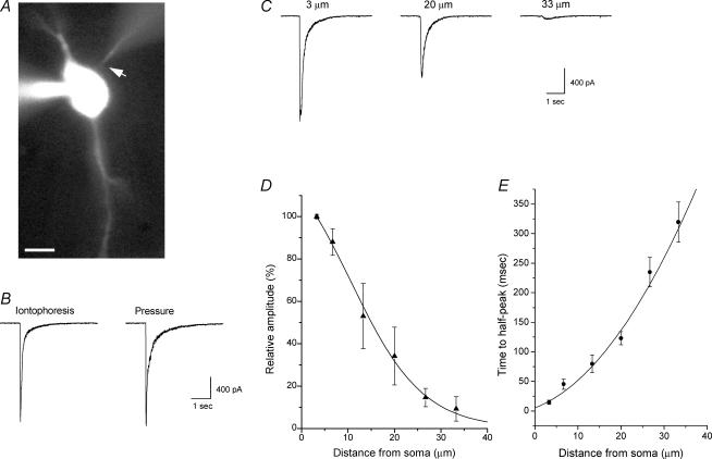
Figure 1. Iontophoretic choline application induces rapid and focal activation of α7 nAChRs in rat hippocampal interneurons in the slice.
A, fluorescence image of a patch-clamped rat CA1 interneuron from the stratum oriens in an acute hippocampal slice. Both the patch and iontophoretic electrodes contained Alexa Fluor 488 (20 μm and 100 μm, respectively) and can be visualized; the iontophoretic electrode is located in close proximity to the soma (arrow). The scale bar is 10 μm. B, brief iontophoretic choline application (100 ms, 200 nA, 1 m choline in iontophoretic electrode) and pressure application of choline (10 mm, 100 ms, right trace) near the soma (∼3–5 μm; left trace) induced similar rapid inward currents in the same cell. C, the amplitude and kinetics of the inward currents activated by the iontophoretic application of choline were dependent on the distance between the tip of the iontophoretic electrode and the soma. The relative amplitude (as a percentage of the response amplitude at a distance of 3 μm) of the responses at variable distances is plotted in D (5 cells), and the data have been fitted to a Gaussian function. The time required to activate responses at variable distances is plotted in E; values plotted are the elapsed time from the initiation of the iontophoretic pulse until the current reached half of the peak amplitude (i.e. time to half-peak).

