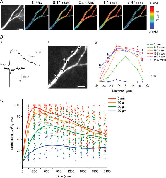
Figure 6. Iontophoretic activation of α7 nAChRs in cultured hippocampal neurons induces [Ca2+]i signals similar to interneurons in slices.
A, the iontophoretic application of choline to the dendrite of a cultured hippocampal neuron (fluorescence image on left shows the location of iontophoretic electrode by dotted lines; the scale bar is 10 μm) induced an increase in [Ca2+]i levels as demonstrated by the pseudo-colour images. B, choline application induced a fast-activating and decaying inward current (bottom trace) and an increase in [Ca2+]i levels (top trace; i); the [Ca2+]i signal was from the region nearest to the iontophoretic electrode (i.e. ‘d’ from the image in ii). The time-dependent changes in [Ca2+]i levels from six different locations for the dendrite (see ii) are shown in iii. C, summary of data obtained from similar experiments at 24 different locations in 5 cells. The changes in [Ca2+]i levels for dendritic regions at variable distances (i.e. 0, 10, 20 and 30 μm) away from the tip of the iontophoretic electrode were plotted as a percentage of the maximal amplitude for the point nearest to the iontophoretic electrode (i.e. 0 μm) versus time after the iontophoretic application of choline. The data for each dendritic location from all experiments were fitted with polynomial functions (5–7 order). The scale bars in A and Bii are 10 μm.

