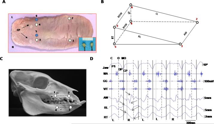
Fig. 1.
– Somomicrometric configuration. (A): crystal implantation. Numbers indicate crystal sites (circles). CP: circumvallate papillae; R and L: right and left. Inset: a pair of piezo ultrasonic crystals. (B): wedge-shaped volume circumscribed by six crystals. AW: anterior width (1–2); BDW and BVW: base dorsal and ventral widths (3–4 and 5–6); RL and LL: right and left body lengths (1–3 and 2–4); RT and LT: right and left base thicknesses (3–5 and 4–6). (C): fluorescent markers (4 dots) and segments (2 lines). (D): synchronized raw recordings of jaw movement (top Ch.), EMG activity (middle 3 Chs.) and tongue deformation (bottom 3 Chs.). Dotted, dashed and solid vertical lines indicate the end of closing (C), start of opening (O) and maximal opening (MO). PS, OP and CP present power stroke, opening and closing phases of jaw movement. L and R indicate chewing side. Solid and dotted arrows indicate peak and valley, respectively, of tongue dimensional changes. Jaw: jaw movement; MA: masseter, GG: genioglossus; V/T verticalis/transversus; AW: anterior width; RL: right body length; RT: right base thickness.

