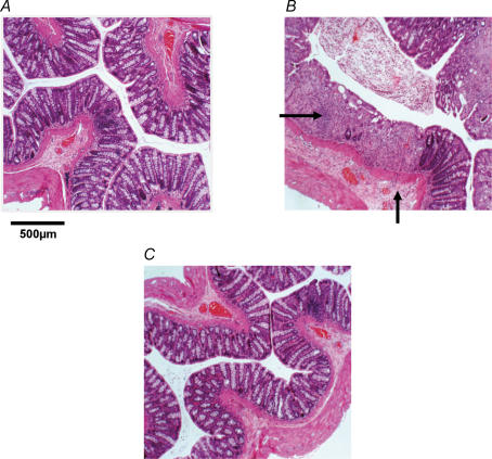
Figure 1. Examples of disease progression and regression in haematoxylin and eosin-stained colonic sections (20 μm).
A, control with normal colonic morphology. B, inflamed colon of a dextran sulphate sodium (DSS)-treated (7 days) animal. The field of view shows examples of crypt destruction, ulceration, oedema and polymorph infiltration in the mucosa and submucosa (arrows), whereas outer muscle and serosa appear unchanged. C, animal 21 days after DSS treatment exhibiting recovery to morphology as in controls.

