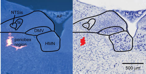
Figure 2. Photomicrographs of a muscimol injection site in the periobex region.
Left, fluorescence photomicrograph showing latex beads. Some beads entered a blood vessel branching medially. Right, brightfield photomicrograph following Nissl stain of the same section. Red area was digitally added to show injection site. AP, area postrema; DMV, dorsal motor nucleus of the vagus; HMN, hypoglossal motor nucleus; NTSis interstitial subnucleus of the nucleus of the solitary tract; ts, solitary tract.

