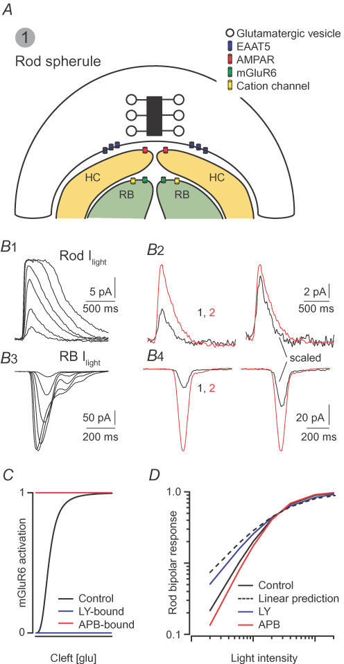
Figure 2. Saturation of postsynaptic glutamate receptor-mediated signalling underlies non-linear signal transfer at the rod–rod bipolar cell synapse.
A, the rod synapse: four postsynaptic processes are located within an invagination having a volume of ∼0.2 μm3 (Rao-Mirotznik et al. 1995). AMPARs on horizontal cell (HC) dendrites are apposed to release sites. Rod bipolar (RB) dendrites are located up to hundreds of nanometres from the presynaptic membrane. Their mGluR6-containing receptors sense a lower [Glu] than the AMPARs. B, light responses from rods and rod bipolars: B1, currents elicited in rods by light flashes (10 ms) of increasing intensity (beginning at 0.75 Rh*, doubling with each successive flash). B2, at left, currents elicited by 0.75 Rh* (black) and 1.5 Rh* (red); scaled responses at right: the response increases linearly with the number of Rh*. B3, currents elicited in rod bipolars by flashes of increasing intensity (beginning at 0.5 Rh*, doubling with each successive flash). B4, left: currents elicited by 0.5 Rh* (black) and 1 Rh* (red); scaled traces at right: doubling the flash strength produces a supra-linear increase in the rod bipolar current. C, schematic illustration of the experiments utilizing mGluR6 agonists and antagonists: LY341495 (antagonist) divides postsynaptic mGluR6 into unoccupied (available to bind glutamate and signal) and inactivated (bound to LY) populations. APB (agonist) also divides the receptors into two populations: unoccupied and activated constitutively (bound to APB). D, LY makes rod bipolar responses more linear, and APB amplifies the non-linearity. F. Rieke provided the traces in B; C and D are modelled on data presented by Sampath & Rieke (2004).

