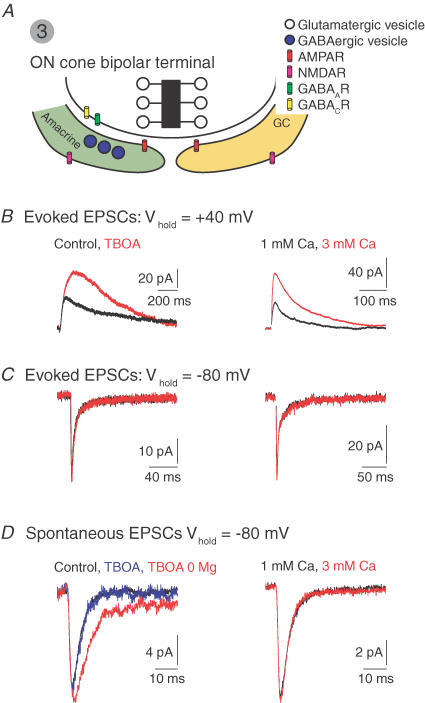
Figure 4. MVR at the ON cone bipolar–ganglion cell synapse.
A, the ON cone bipolar synapse. Postsynaptic amacrine and GCs express synaptic AMPARs and peri-synaptic NMDARs. The amacrine makes a reciprocal GABAergic synapse with the terminal. B, blocking glutamate uptake with DL-threo-β-benzyloxyaspartate (TBOA, left) or increasing PR (elevating external [Ca2+], right), potentiates evoked NMDAR- but not AMPAR-mediated EPSCs C. D, spontaneous AMPAR-mediated EPSCs are unaffected by TBOA and elevated [Ca2+]. In the absence of Mg2+ (permitting NMDARs activation at negative potentials), TBOA induces an NMDAR-mediated component of the sEPSC. 0 Mg2+ alone does not reveal an NMDAR-mediated component. J. Diamond provided the illustrated traces.

