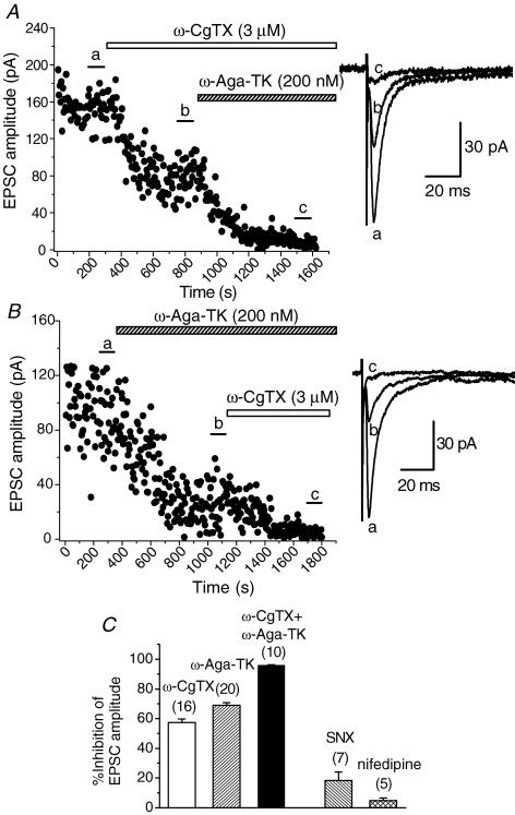
Figure 5. Suppression of the EPSCs by ω-CgTX and ω-Aga-TK.
A and B, time course of the inhibitory effects of ω-CgTX (3 μm, A) and ω-Aga-TK (200 nm, B) on the amplitude of EPSCs. Toxins were bath applied during the indicated periods. A,ω-CgTX reduced the amplitude of EPSCs by 49.0% of control. B,ω-Aga-TK reduced the amplitude of EPSCs by 66.3% of control. Subsequent application of ω-Aga-TK (A) or ω-CgTX (B) almost blocked the remaining EPSCs. Superimposed sample records at the right of A and B are the averages of 20 consecutive EPSCs during the indicated periods. C, histograms summarizing the suppression of the EPSCs by ω-CgTX (3 μm), ω-Aga-TK (200 nm), SNX-482 (SNX, 300 nm) and nifedipine (10 μm). Values for ω-CgTX, ω-Aga-TK, both ω-CgTX and ω-Aga-TK, SNX and nifedipine are 57.5 ± 2.27% (n = 16), 68.9 ± 1.71% (n = 20), 96.1 ± 0.27% (n = 10), 18.3 ± 5.70 (n = 7) and 4.70 ± 1.80 (n = 5), respectively.

