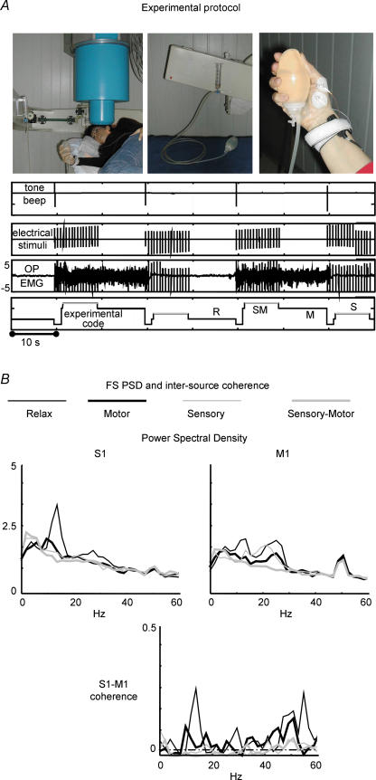
Figure 6. Sensorimotor feedback.
A, experimental protocol: Top from left: (1) subject position during recordings from scalp positions overlying the peri-rolandic area contra-lateral to the moved-stimulated hand: (2) non-magnetic device, i.e. the water sphygmomanometer, to control the level of contraction: (3) showing the thumb position during isometric contraction, with electrodes recording OP EMG signal, as well as the median nerve stimulation at wrist. Bottom from top: (1) trigger indicating tone beep for starting/stopping isometric contraction: (2) trigger indicating sensory stimuli to the median nerve at wrist: (3) electromyographic signal, where relax and contraction periods are clearly noticeable, as well as the sensory stimuli artifact: (4) off-line generated signal code differentiating the 4 experimental conditions: relax (R), sensory (S), motor (M), sensory-motor (SM). B, FS PSD and intersource coherence. PSD of the S1 and M1 cerebral sources (top) and S1–M1 coherence (bottom) in the four experimental conditions in a representative subject. Note: higher S1 representation in the alpha band and higher M1 beta and gamma band representation; lower S1–M1 coherence in all condition with respect to rest in the three frequency bands and values above significance threshold in motor condition (dashed–dotted line). Note that the y axes of S1 and M1 PSDs do not have measurable units, as FSs do not have a physical unit dimension. The S1–M1 coherence is a dimensionless quantity.

