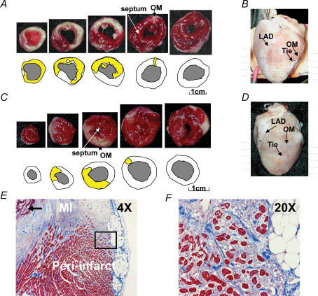
Figure 1. Two types of subacute myocardial infarction.
A and C, show the results of triphenyl tetrazolium chloride staining and the methods used to determine infarct size. The sections were taken at sites shown by the horizontal lines in B and D. E, shows low power (4 ×) view of trichrome-stained peri-infarct zone showing transmural myocardial infarction and adjacent peri-infarct zone. Residual necrotic, ‘mummified’ myocardium in the centre of the infarct is seen in the left upper corner of the field (arrow). F, shows a higher power (20 ×) view of the field within the black square in E, demonstrating surviving myocytes (red) surrounded by fibrous tissue (blue). OM, obtuse marginal branch of left circumflex artery; LAD, left anterior descending artery; MI, myocardial infarction.

