Figure 6.
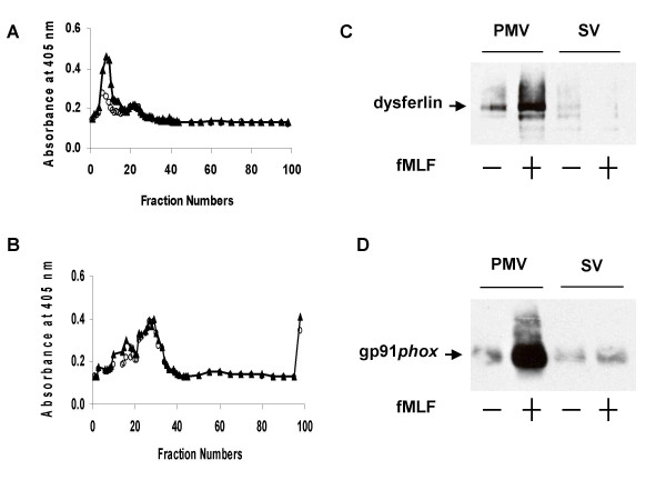
Secretogogue-induced redistribution of dysferlin. (A) Isolated resting PMN were disrupted by N2 cavitation and fractionated using a two-step discontinuous gradient of Percoll. The γ band containing the light membranes was recovered, treated with neuraminidase, and subjected to free-flow electrophoresis to resolve plasma membranes vesicles from secretory vesicles. Fractions (96) were collected and assayed spectrophotometrically for alkaline phosphatase activity in the absence (○) and presence (△) of Triton X-100. (B) The PMN were isolated as above and were exposed to fMLF (formyl methionyl-leucyl-phenylalanine). Fractions (96) were collected and assayed spectrophotometrically for alkaline phosphatase activity in the absence of (○) and presence (△) of Triton X-100 after exposure to fMLF. The exposure to fMLF resulted in a loss of SV (i.e. loss of latent alkaline phosphatase activity), consistent with their fusion with the plasma membrane. (C) Purified PMV and SV from resting or fMLF-stimulated PMN were separated by SDS-PAGE, electroblotted, and the resulting blots probed with anti-dysferlin. (D) As a control for intracellular membrane recruitment, samples were also probed with 54.1, as flavocytochrome b558 expression at the PMN surface increases with agonist-stimulated granule and secretory vesicle fusion with plasma membrane.

