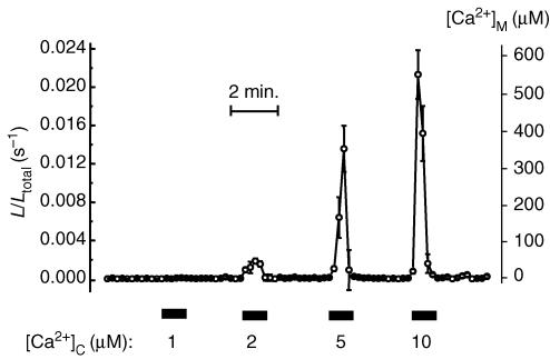
Figure 2. Ca2+ dependence of mitochondrial Ca2+ uptake in permeabilized sympathetic neurons.
Sympathetic neurons infected with mitmutAEQ and reconstituted with coelenterazine n were permeabilized by treatment with 20 μm digitonin over 1 min at 37°C in Ca2+-free (containing 0.2 mm EGTA) intracellular-like medium (composition, in mm: NaCl, 5; KCl, 130; MgCl2, 2; KH2PO4, 2; Mg-ATP, 0.2; and potassium-Hepes, 20; pH 7.0 adjusted with KOH). Then the cells were perfused with ‘intracellular-like’ medium containing different [Ca2+] values, as shown in the figure. The different [Ca2+] values were obtained using buffers containing 1 mm EGTA or 1 mm N-(hydroxyethyl) ethylenediamine triacetic acid (HEDTA) and adjusting Ca2+ and Mg2+ as required (Patton et al. 2004). Values represent the mean ± s.e.m. of 5 different cells present in the same microscopic field. Results are representative of 5 similar experiments.

