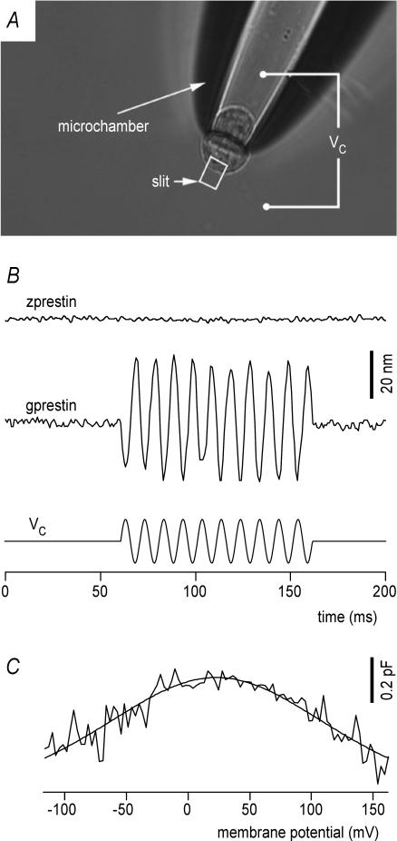
Figure 4. zprestin-transfected cells do not display somatic electromotility.
A, transfected HEK cells were probed for electromotility in a microchamber configuration, which allows simultaneous electrical stimulation and displacement recording. The rectangular slit indicates the area that was projected onto a photodiode for motility measurements. The stimulus command voltage (Vc) was applied between bath and microchamber compartment. Proportional to their degree of insertion into the microchamber, the cell's resultant membrane voltage swing was calculated to be ∼60% of Vc. B, lack of somatic motility in a zprestin-transfected HEK cell (upper trace). A 380 mV (peak-to-peak) voltage burst (lower trace) was applied via the microchamber. The voltage drop on the extruded segment was estimated to be 228 mV (peak-to-peak). The cell's resting membrane potentials normally were around −10 mV. Therefore, the effective membrane voltage was estimated to vary from −124 to +104 mV. Note that in contrast, robust electromotiltiy was observed in gprestin-transfected cells (middle trace). C, HEK cells transfected with zprestin display NLC confirming proper membrane targeting of zprestin in HEK cells. Voltage-dependent capacitance was measured using a 2-sine-wave method.

