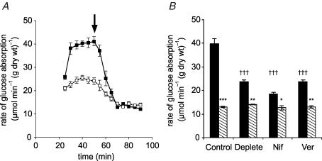
Figure 1. The effect of luminal Ca2+ and L-type effectors on glucose absorption.
A, a representative time course showing the effect of Ca2+. Rat mid and distal jejunum was pre-perfused in vivo in single-pass mode with modified KHB + 100 mm mannitol containing either 1.25 mm Ca2+ (▪) or no Ca2+ (□, Ca2+-deplete perfusate); after 20 min the perfusate was switched to 75 mm glucose + 25 mm mannitol to achieve an initial steady state (30–50 min) before the addition of 1 mm phloretin at 50 min (arrow) to determine the phloretin-insensitive (SGLT1) steady-state rate (60–90 min). B, the effect of L-type effectors, which were present from the start of perfusions. Total (black bars) and phloretin-insensitive (hatched bars) steady-state rates were obtained for control, Ca2+ deplete, nifedipine (10 μm, Nif) and verapamil (100 μm, Ver) perfusions (n=4, each case). Rates are means ± s.e.m. expressed in μmol min−1 (g dry wt)−1. Student's t test was used to determine statistical significance. †††P < 0.001, unpaired test for the comparison of the initial steady-state rates of the Ca2+ deplete, nifedipine and verapamil to that of the control. ***P < 0.001, **P < 0.01, *P < 0.05, paired test comparing the initial steady-state rate with its phloretin-insensitive rate. The phloretin-insensitive rates are not statistically different from one another.

