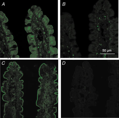
Figure 7. Immunocytochemical localization of Cav1.3 and β3-subunit in the proximal jejunum.
Sections of unperfused jejunum were labelled with a rabbit antibody that recognizes all forms of rat Cav1.3 (A and B) and with a rabbit antibody which specifically recognizes the β3 subunit (C and D). The secondary antibody was FITC-conjugated goat anti-rabbit IgG. The peptide controls were sections treated with antibody to Cav1.3 (B) and β3 (D) that had been pre-absorbed with excess antigenic peptide; non-specific staining in the lamina propria of these sections was seen with the secondary alone. All sections are at 63 × magnification and were taken at the same settings with a Zeiss LSM 510 confocal microscope.

