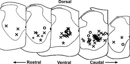
Figure 2. Location of recorded cells.
The locations of 67 of the recorded cells were estimated from postrecording digital pictures of electrode tip position (see Methods, cell identification). Rostral to caudal locations are shown left to right; dorsal is to the top and ventral is to the bottom. The medial side of the slice half is indicated with a vertical line on the right side of each slice, with the central canal shown as a darkened half oval. Firing patterns of cells are marked as follows: Repetitive-firing: ×; repetitive-burst: ○; initial-burst: +'; single-spiking: *.

