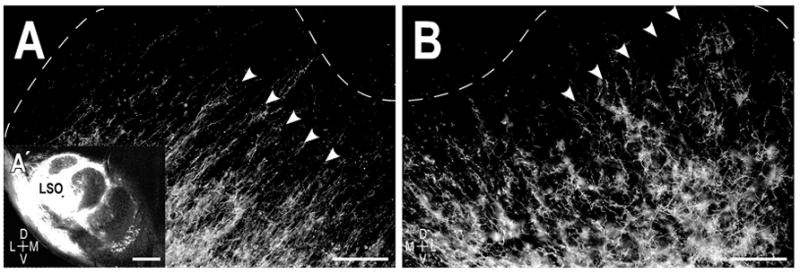
Figure 2.

Resultant afferent labeling in the central nucleus of the IC in a P0 cat. A. Axonal layers (arrowheads) in the left IC ipsilateral to the LSO dye placement (A′ inset). B. Labeled axons in the contralateral IC exhibited a similar laminar bias, forming a periodic pattern of afferent bands (arrowheads) that spanned the frequency axis of the central nucleus. Dashed contours delineate boundaries of the central nucleus with the dorsal cortex and the external cortex as determined by the counterstaining. Scale bars A, B = 200 μm, A′ = 500 μm.
