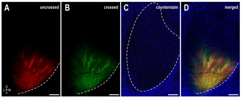
Figure 6.

Interdigitation of crossed and uncrossed LSO axonal layers in the left IC at P0. A. DiI labeling illustrating ipsilateral LSO bands. B. Same field as (A) showing DiD label arising from the contralateral LSO. C. Pseudocolored image of a Nissl stain of the same field of view highlighting boundaries of the central nucleus (dashed contour). D. Digitally merged illustration of (A – C) showing the spatial relationship of uncrossed and crossed LSO projection domains within the central nucleus of the IC. Dashed contours in A, B, and D depict the ventromedial border of the IC. Scale bars = 500 μm.
