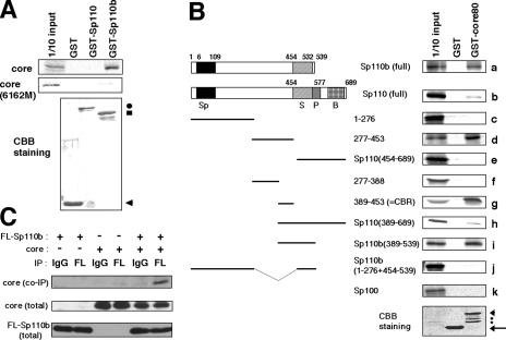
FIG. 3.
Interaction of the core with Sp110b. (A) The 35S-labeled in vitro transcription-translation product of full-length core (top panel) or the core(6162M) mutant (center panel) was incubated with recombinant Sp110 or Sp110b fused to GST (GST-Sp110 or GST-Sp110b) or with GST as a negative control. The GST pulldown assay was performed as described in Materials and Methods. 1/10 input, signal for 1/10 the amount of 35S-labeled product used in the pulldown assay. Coomassie brilliant blue (CBB) staining patterns of pulled-down proteins are shown in the bottom panel. Arrowhead, circle, and square indicate the bands corresponding to GST, GST-Sp110, and GST-Sp110b, respectively. (B) Mapping of the region interacting with the core in Sp110b by deletion analysis. (Left) Schematic representations of the full-length and truncated mutants of Sp110 and Sp110b are shown. Numbers above the diagrams indicate the amino acid positions from the amino terminus of Sp110 or Sp110b. The Sp100-like domain (Sp), SAND domain (S), PHD (P), and bromodomain (B) are indicated. (Right) Designations of the GST fusion protein and 35S-labeled derivatives of Sp110 and Sp110b are given above and to the left of the autoradiograms (a through k), respectively. GST-core80, GST fused with the region of the core from aa 1 to 80. CBB staining patterns of pulled-down proteins are shown in the bottom panel. Arrow and arrowhead indicate bands corresponding to GST and GST-core80, respectively. Two dots indicate apparent degradation products of GST-core80. (C) Interaction between the core and Sp110b produced in the cells. Lysates from COS-7 cells transfected with 1 μg of pCMV-Sp110b and/or pCMV-core were used for coimmunoprecipitation, followed by immunoblot analysis. The combinations of plasmids used for the transfection are indicated at the top. “IP” designates the antibodies used for immunoprecipitation, either the anti-FLAG antibody (FL) or normal mouse IgG (used as a negative control). Coimmunoprecipitated core with FLAG-tagged Sp110b was detected with an anti-core antibody (top panel). Center and bottom panels show results of experiments in which the core and FLAG-tagged Sp110b in total-cell lysates from transfectants were detected by anti-core and anti-FLAG antibodies, respectively.

