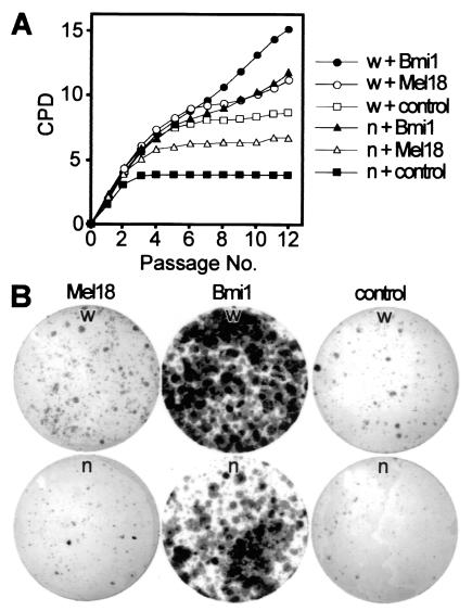
FIG. 8.
Growth properties of Cited2−/− and Cited2+/+ embryonic fibroblasts infected with Bmi1- and Mel18-expressing retroviruses. (A) Proliferation of Cited2−/− (n) and Cited2+/+ (w) fibroblasts (derived from littermate embryos on a C57BL/6J background at 13.5 dpc) infected with Bmi1, Mel18, or control retroviruses. Fibroblasts were harvested, infected with retroviruses the following day, replated on day 4 (referred to as P0), and passaged in parallel every 3 days at identical conditions. The proliferation of retrovirally complemented fibroblasts is shown as plots of CPD versus passage number. (B) Colony formation assay. Cited2−/− and Cited2+/+ fibroblasts (at passage 6) infected with the indicated retroviruses were plated at a density of 4,000 cells per 9-cm plate, and colonies were visualized with Giemsa stain after 16 days.

