FIG. 1.
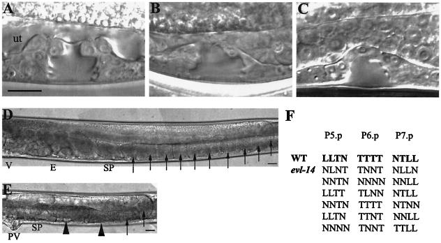
Vulval and gonadal abnormalities caused by evl-14(ar96) and scc-3(ku263). All images are oriented with the anterior portion to the left and the dorsal portion to the top. (A to C) Mid-L4-stage vulva in a wild-type and two mutant animals. (A) The wild-type vulva (containing 22 nuclei) displayed a symmetric “Christmas tree” shape of the vulval lumen. “ut” indicates the location of the uterine space. (B and C) The irregular and asymmetric shape of the vulval lumen displayed in evl-14 (B) and scc-3 (C) mutants resulted from fewer vulval cells. (D) Posterior arm of the gonad in a young adult wild type. V, vulva; E, embryos; SP, spermatheca. Arrows indicate oocytes. (E) Posterior arm of the gonad in a young adult evl-14 mutant animal. Note lack of embryos and displayed endomitotic oocytes. PV, protruding vulva. Arrowheads indicate endomitotic oocytes. Similar gonad phenotypes were seen in scc-3 mutants. (F) Lineage analysis of the evl-14 mutant vulva. Each line represents either a wild-type (first line) or evl-14 mutant pattern of final divisions of P5.p to P7.p (23). T, cell division along the left-right (transverse) axis; L, cell divided along the anterior-posterior (longitudinal) axis; N, cell did not divide. The genotypes of the two mutant strains used for the experiments whose results are shown here and in Fig. 4 to 6 are as follows: unc-36(e251) evl-14(ar96) and rol-4(sc8) scc-3(ku263). Bars, 10 μm.

