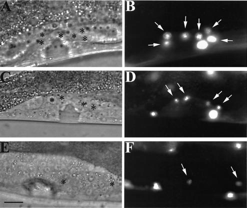
FIG. 2.
scc-3 and evl-14/pds-5 mutants have abnormal π cell linage. (A, C, and E) Differential interference contrast images of L4 hermaphrodites carrying an integrated cog-2::gfp fusion construct. (B, D, and F) Fluorescence images corresponding to images in panels A, C, and E. Arrows, π cells. (A and B) Wild-type worm; (C and D) evl-14 RNAi-treated worm; (E and F) scc-3 RNAi-treated worm. In the fluorescence images, cog-2::gfp expressed in π cells and body wall muscle cells is depicted. Asterisks in panels A, C, and E indicate π cells. Six π cells (ventral uterine cells) are visible in the wild-type image (B). Fewer π cells appeared in the animals treated with either evl-14 RNAi or scc-3 RNAi. Bar, 10 μm.

