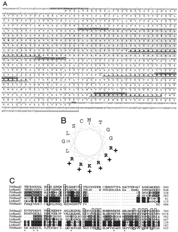
FIG. 1.
Band II sequence. (A) Upper line, the nucleotide sequence (the longest cDNA, plus in parentheses the RT-PCR-determined 5′ end). Oligonucleotide primers are indicated in boldface and uppercase (pairs have common underlining). The dsRNA region, start and stop codons, and zinc finger are indicated by highlighting. Lower line, the protein sequence. The three sequenced tryptic fragments are boldface amino acids; residues that confirmed the initial PCR fragment are singly underlined, and residues that additionally confirmed the cDNA are doubly underlined. (B) Helical wheel representation of the first 15 amino acids, indicating an amphipathic alpha helix. (C) C-terminal region of T. brucei band II (TbMP81), compared with the analogous region of T. brucei band VI (TbMP52), VII (TbMP18), and III (TbMP63) proteins and to L. major homologs by use of T-Coffee (27). Highlighting indicates cross-species conservation. Boxes and stars show identity and charge conservation, respectively between the different proteins. The L. major band III homolog is not included, as it lacks part of this C-terminal region.

