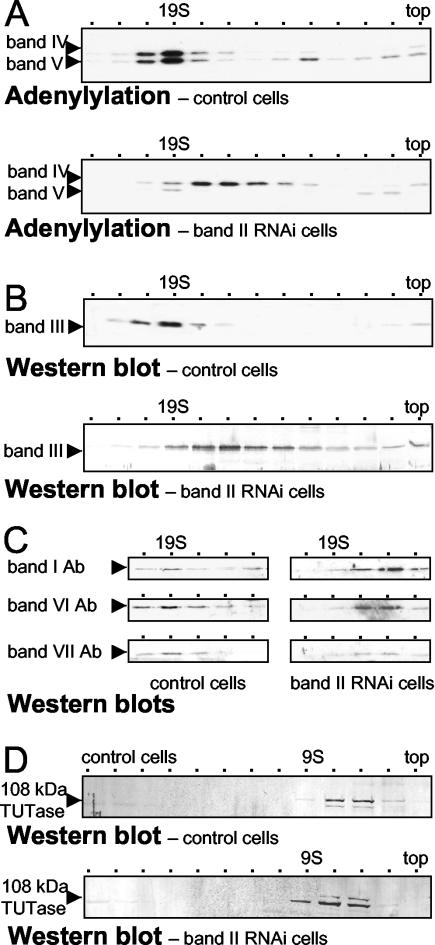
FIG. 5.
Sedimentation analysis of editing components from band II RNA cells. In parallel, mitochondrial extracts were prepared from a clonal band II RNA cell line and 29.13 control cells at 5 days postinduction and were resolved by glycerol gradient centrifugation. (A) Adenylylation assays of fractions 4 to 16 (top) from the control cells (upper) and band II RNAi cells (lower), following deadenylylation. ∼19S peaks in fraction 7. (B) Fractions 4 to 16 (top) from the control cells (upper) and band II RNAi cells (lower) were detected by using antibody to band III protein. (C) Fractions 7 to 10 from the control cells (left) and band II RNAi cells (right), including the ∼20S and ∼15S regions, were analyzed by using antibodies (Ab) to each of the other major proteins of the basic editing complex. (D) Fractions 4 to 16 (top) from the control cells (above) and band II RNAi cells (below) were detected by using antibody to the 108-kDa TUTase. Because this TUTase near the top fraction (last fraction collected) had different volumes in these two gradient, it appears in slightly shifted fractions.

