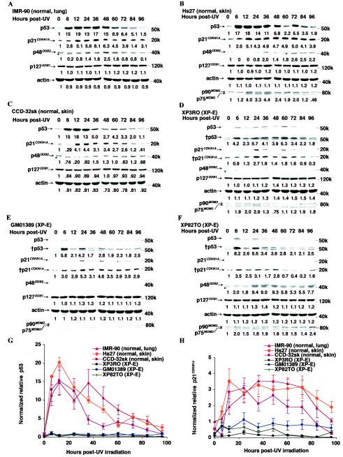
FIG.3.
p53 levels after UV irradiation are severely reduced in XP-E strains. (A to F) Protein levels of p53, p21CDKN1A, p48DDB2, p127DDB1, and actin after 12 J of UV irradiation m−2 in normal (A to C) and XP-E (D to F) strains. The chemiluminescence exposure times were 20 s (p53), 2 min (†p53), 15 s (p21CDKN1A), 3 min (†p21CDKN1A), 20 min (p48DDB2), or 10 s (MDM2, p127DDB1, and actin). The band marked with an asterisk lying just below p48DDB2 is nonspecific. Each basal protein level (0 h) was designated as 1.0 to compare protein levels during the time course. (G and H) Quantitation of p53 (G) and p21CDKN1A (H). All protein levels were normalized of the protein levels in the IMR-90 cells at 0 h after being normalized for the amount of actin. Each value was determined by six (XP3RO), four (CCD-32sk and GM01389), three (IMR-90 and Hs27), or two (XP82TO) experiments for p53 levels; six (Hs 27), three (IMR-90, CCD-32sk, GM01389, and XP82TO), or two (XP3RO) experiments for p21CDKN1A levels.

