FIG. 2.
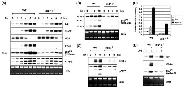
Dependence of UPR target gene expression on XBP-1. (A) wt and XBP-1−/− MEF cells were treated with 10 μg of Tm/ml for the indicated time periods. Total RNAs were isolated and subjected to Northern blot analysis. The same blot was hybridized sequentially with BiP, CHOP, MGP, ERdj4, p58IPK, ATF6α, and MGP probes. The p58IPK probe is probe A, from the 3′ end of the gene. Ethidium bromide staining of the gel before blotting is shown at the bottom for loading control. (B) All isoforms of p58IPK are XBP-1 dependent. Here, Northern blot analysis was performed with probe B, which recognizes sequences at the 5′ end of the gene. (C) XBP-1-dependent genes are also IRE1α dependent. Northern blot analysis of RNA prepared from IRE1α−/− MEFs treated with Tm for various time periods and assessed for expression of ERdj4 and p58IPK. (D) The ERdj4GL3 reporter was transfected with or without XBP-1s plasmid into wt and XBP-1−/− MEF cells. Cells were treated with Tm at 1 μg/ml for 16 h before harvesting as indicated. Luciferase activity was normalized to the Renilla activity. (E) Induction of BiP, ERdj4, and p58IPK in primary B cells by LPS. B220+ primary B cells were isolated from spleens of wt or XBP-1−/− RAG2−/− lymphoid chimeras. Cells were untreated or stimulated for 3 days with 20 μg of LPS/ml. Expression of BiP, ERdj4, and p58IPK was determined by Northern blot analysis.

