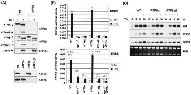
FIG. 4.
UPR target gene expression is largely unaffected in the absence of ATF6α and ATF6β. (A) Western blot analysis of iATF6α-, iATF6β-, and double iATF6α/β-expressing MEFs. MEF-iATF6α cells were generated by transfecting MEF cells with U6-iATF6α plasmid, which expresses siRNA for ATF6α under the control of the U6 promoter. MEF-iATF6β and MEF-iATF6α/β cells were generated by transducing wt and MEF-iATF6α cells with retroviruses that express iATF6β. Lysates from wt and MEF-iATF6α, MEF-iATF6β, and MEF-iATF6α/β MEFs either not treated or treated with 10 μg of Tm/ml for 6 h were analyzed for the expression of XBP-1, ATF6α, and ATF6β. An asterisk indicates a nonspecific band recognized by anti-ATF6α antibody. (B) 5xATF6GL3 (UPRE) or ERSE reporters were transfected into wt, XBP-1−/−, iATF6α, iATF6β, double iATF6α/β, and double XBP-1−/−/iATF6α MEF cells. Cells were treated with Tm at 1 μg/ml for 16 h before harvesting as indicated. Luciferase activity was normalized to Renilla activity. The fold induction of relative luciferase activity by Tm treatment compared to untreated samples is also shown. (C) Total RNA was prepared to measure the expression level of BiP, CHOP, ERdj4, p58IPK, and Grp94 mRNAs. Ethidium bromide staining of the gel before blotting is shown at the bottom.

