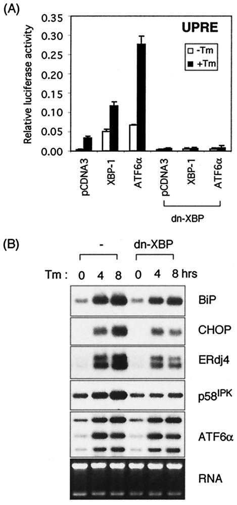
FIG. 6.
Dominant negative XBP-1 suppresses both XBP-1 and ATF6α activity. (A) The 5xATF6GL3 reporter plasmid was cotransfected with either pCGNATF6α or XBP-u/s plasmids into MEF cells with or without the dominant-negative XBP-1 expression plasmid. A total of 100 ng of DNA was used for each transfection except for pCDNA3.1, which was added to give 1 μg of DNA in total. Luciferase assays were performed as described in the legend to Fig. 4. (B) MEF and MEF-dn-XBP cells that stably express dominant-negative XBP-1 protein were treated with 10 μg of Tm/ml for the indicated time periods. Total RNAs were isolated and subjected to Northern blot analysis. The same blot was hybridized sequentially with BiP, CHOP, ERdj4, p58IPK, and ATF6α probes. Ethidium bromide staining of the gel before blotting is shown at the bottom as a loading control.

