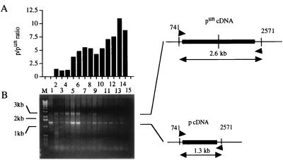
Figure 2.
Detection of wt p mRNA in black spots on the gray coat of pun/un mice. Black spots and similar gray areas were excised from 3- to 5-day-old mice delivered by treated and control dams. (A) The RT-PCR analysis was performed using primers spanning duplicated regions. The samples were run in 1% agarose gels and stained with ethidium bromide. (B) Photographs of stained gels were analyzed by scanning densitometry with a BioImage (Millipore). The relative intensity of the wt p mRNA was evaluated as the ratio of bands’ intensities corresponding to p and pun. Lanes: M, 1kb DNA ladder; 1, wt black p/p mouse; 2, gray pun/un mouse; 3 and 4, gray skin from x-ray-treated pun/un mice; 5–7, black spots from x-ray-treated pun/un mice; 8–10, black spots from EMS-treated pun/un mice; 11 and 12, black spots from SOA-treated pun/un mice; 13 and 14, black spots from BEN-treated pun/un mice; 15, control RT-PCR without RNA.

