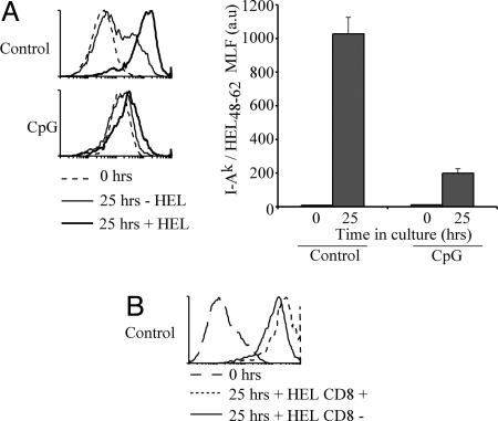
Fig. 2.
Mature DCs are no longer able to present newly encountered antigens. (A) Purified DCs from control (Upper) or CpG-pretreated (Lower) CBA mice were incubated with or without 1 mg/ml HEL for 25 h and stained with the mAb AW3.1. The accumulation of I-Ak-HEL48–62 was assessed by flow cytometry. Representative flow cytometry profiles from two experiments are shown. The dashed line shows the staining level before incubation. The bar graph (Right) displays the MLF values of samples incubated with HEL after subtraction of the values in the corresponding samples incubated without HEL. The result represents the mean of duplicate samples; error bars indicate SD. The results shown are representative of two experiments. (B) As in A, but using segregated CD8+ and CD8− DCs purified from control mice. The result is representative of two experiments with each sample performed in duplicate.

