Fig. 2.
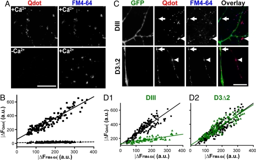
Qdot uptake is Ca2+-dependent and clathrin-mediated. (A) (Left) Qdot loaded in the presence (Upper) or absence (Lower) of Ca2+. (Right) FM4-64 loaded in the same fields (4× 10-Hz 2-min stimulation) in the presence of Ca2+. (Scale bar, 10 μm.) (B) The relationship between Qdot loading (|ΔFQdot|) and FM4-64 staining (|ΔFFM4-64|) is significantly different depending on the presence of Ca2+ during loading (squares) or the absence (triangles) (P < 0.001, Kolmogorov–Smirnov test). Each data point represents an individual ROI. (C) EGFP fluorescence reveals neurons expressing DIII-EGFP or D3Δ2-EGFP, and retrospective FM4-64 staining marks functional boutons. Boutons of DIII–EGFP expressing neurons (arrow) take up Qdots to a significantly lesser degree than those of nontransfected neurons (arrowhead). Boutons of neurons expressing D3Δ2-EGFP (arrow) take up Qdots similarly to those of nontransfected neurons (arrow head). (Scale bar, 5 μm.) (D1 and D2) The relationship between Qdot loading (|ΔF Qdot|) and FM4-64 staining (|ΔF FM4-64|) is significantly different for DIII-EGFP expressing neurons (green triangles) and nonexpressing neurons in the same culture (black squares) (D1, P < 0.001, Kolmogorov–Smirnov test). Expression of the inactive D3Δ2-EGFP (green triangles) gives no significant difference from nonexpressing control neurons (black squares) (D2, P > 0.5, Kolmogorov–Smirnov test). Note that FM4-64 staining was spared under all circumstances, likely because 4× high K+ stimulation was greatly supermaximal for loading and dye could load even with K&R.

