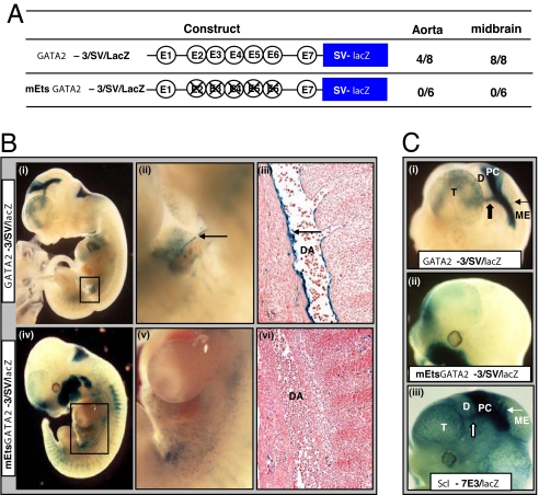
Fig. 4.
The Gata2-3 enhancer targets the hemogenic aortic endothelium and requires Ets sites for its in vivo activity. (A) Transgenic lacZ reporter constructs with WT enhancer (Gata2-3) or enhancer with mutant Ets sites (mEts Gata2-3). The number of embryos showing staining in the DA (AGM region) or midbrain out of the total number of E10.5 and E11.5 F0 transgenic embryos generated with each construct is listed alongside. (B) Representative F0 embryos stained with X-Gal for lacZ expression. (i) Gata2-3/SV/lacZ E10.5 whole-mount showing staining in the DA (boxed). (ii) Magnified view of the boxed area in i showing concentration of the reporter along the ventral surface of the DA (arrow). (iii) Histological section corresponding to region in (ii) showing prominent endothelial staining (arrow). (iv) mEts Gata2-3/SV/lacZ E11.5 whole-mount embryo showing nonspecific staining in the region of the DA (boxed). (v) A magnified view of the boxed area in iv. (vi) Section corresponding to region in v showing absence of endothelial staining. (C) Magnified lateral view of heads of X-Gal stained F0 embryos in B. (i) Gata2-3/SV/lacZ whole-mount embryo showing prominent lacZ expression in the posterior commissure (block arrow) and tectal neurons into the mesencephalon (arrow). (ii) mEts Gata2-3/SV/lacZ whole-mount embryo showing loss of lacZ staining. The ectopic staining in the maxillary region was seen in only one of six F0 mutant transgenic embryos. (iii) Scl-7E3/lacZ wholemount embryo showing staining similar to that seen with the Gata2-3/SV/lacZ transgenics. D, diencephalon; ME, mesencephalon; PC, posterior commissure; T, telencephalon.

