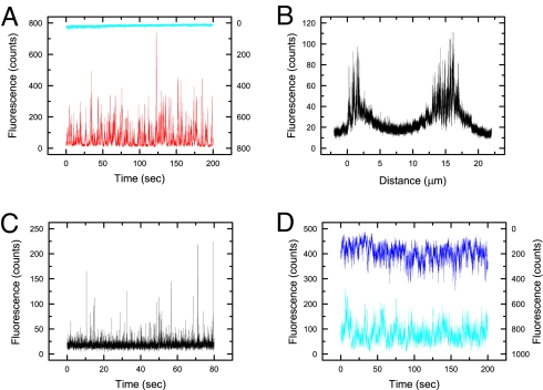
Fig. 2.
Control experiments confirming single-molecule level fluorescence detection. (A) Vβ8-KMAC92.6 cells incubated with Alexa488-labeled anti-TCRβ Fab fragments (nonbinding, blue trace) and anti-CD3ε Fab fragments (binding, red trace). Fluorescence bursts are only observed for the anti-CD3ε Fab, indicating that the bursts result from the binding of the fluorescent Fabs to their target antigens at the cell surface. (B) A DO11.10 cell incubated with Alexa488-labeled anti-TCRβ Fab fragments was scanned across the diameter of the cell by using a scan rate of 1 μm/25 s. Fluorescence bursts are only observed at the perimeter of the cell. (C) A DO11.10 cell stained with the DiO membrane probe; single-molecule fluorescence bursts are observed at the same focus height (15 μm) as the bursts detected in Fig. 2B. (D) Fluorescence from Yae5B3K cells incubated with Alexa647-labeled anti-CD3ε Fab fragments and excited at different laser powers. No differences in the bursts was observed, beyond a 50% reduction in overall fluorescence, for cells illuminated with the laser power routinely used (1 μW; dark-blue trace) versus half that typically used (0.5 μW; blue trace), suggesting that optical trapping effects are absent.

