Fig. 2.
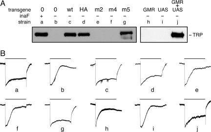
Transgenic rescue of the inaF phenotype. (A) Total extracts of head proteins from various strains were probed with anti-TRP. All strains (except for a) carried the X-linked inaFP106x deletion allele. Strains labeled c–g also carried a 13-kb segment of genomic DNA from the inaF region on an autosome. In strain c, this transgene carried the wild-type sequence; in strain d, the wild-type sequence was kept intact, but an HA tag was appended to the C terminus of the 81-aa ORF; in strains e and f, this ORF was modified by introduction of a triple substitution and a frameshift, respectively; in strain g, the 241-aa ORF on the transgene was modified by introduction of a frameshift. Strains labeled h–j carried, as indicated, a transgene bearing the eye-specific GMR-GAL4 driver (23), a UAS transgene bearing the 3.1-kb inaF cDNA, or both transgenes. Construction of the inaF transgenes is described in Materials and Methods; the sequence alteration of the transgene in strains d–f is given in Fig. 1B. (B) Electroretinograms recorded as described (24) from adults of the strains whose genotype is given in A. The bar above each trace shows the 20-s period during which these dark-adapted flies were exposed to white light (88,000 lux). Each trace was normalized to the potential difference that developed 0.25–0.50 s after the onset of light pulse. In traces a–g and h–j, the absolute value of this potential ranged from 16 to 22 mV and from 12 to 14 mV, respectively. In unrescued or poorly rescued flies (b, e, f, h, and i), by the end of the light pulse the photoreceptor potential decayed to the baseline or close to it. Conversely, in wild-type and well rescued flies (a, c, d, g, and j), the photoreceptor potential is still substantial at the end of the light pulse.

