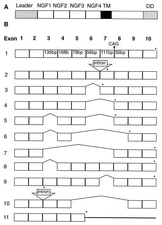
Figure 5.
(A) Schematic representation of the LARD protein drawn above the proposed genomic structure showing the leader peptide, four NGFR repeats, the transmembrane domain, and the death domain. (B) A proposed scheme for the genomic structure of LARD is represented above the prototype LARD-1 cDNA. Alternatively spliced clones LARD-2 through -11 are represented below LARD-1. Positions of the termination codons are represented by an asterisk above the clones. The CAG insertion in clone LARD-1b is indicated above LARD-1.

