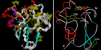
Fig. 3.
The hydrophobic core of Ros87. (Left) Superposition of the best NMR structures (residues 9–66) to show the polypeptide backbone, the four zinc-coordinating residues, and the 15 hydrophobic core side chains. The four zinc-coordinating residues are depicted in magenta, the three corresponding to the eukaryotic hydrophobic core are in cyan, and the others are in yellow. (Right) Three relevant hydrogen bonds (white) forming in Ros87 NMR structure.

