Fig. 4.
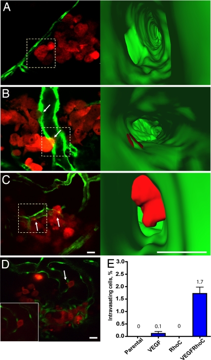
RhoC cooperates with VEGF to enhance tumor cell intravasation. Single optical sections showing tumor cells interacting with the vessel surface (5 dpi). (A–C) MDA-435 expressing RhoC (A), VEGF (B), or RhoC and VEGF (C). (Right) Three-dimensional reconstructions of interior vessel surfaces at the tumor cell-vessel interface (dotted squares in Left). Arrows show vascular mimicry (B) or membrane protrusion into the vessel lumen (C). In C, note the large increase in the membrane protrusion inside the vessel lumen of the MDA-RhoC cells secreting VEGF (SI Movies 8–10). (D) Single optical section showing an MDA-RhoC VEGF membrane protrusion in the vessel lumen (arrow). (Inset) The same cell 5 min later. (E) Plot showing percent of intravasating cells for parental MDA-435 cells or MDA-435 cells expressing VEGF, RhoC, or RhoC and VEGF. MDA-RhoC cells that express VEGF had a significantly higher percentage of intravasating cells (P < 0.05, t test) than other cell types. Mean and SEM values are displayed above the plot. Colors code: Fish vasculature is green, and human tumor cells are red. [Scale bars, 20 (Left) or 10 μm (Right).]

