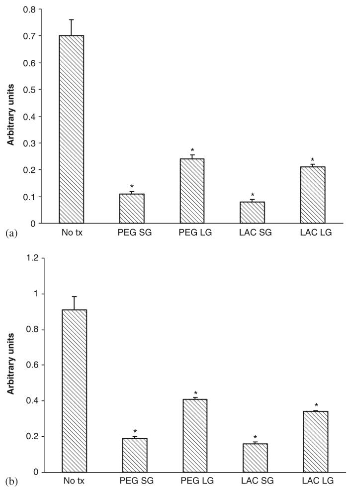
Fig. 6.

Effect of gels on (a) human dermal fibroblast and (b) keratinocyte cell motility. Gels composition was as described in previous figure legends. Cells were exposed to gels after 48 h in serum depleted media and motility assessed by an in vitro wound healing assay. Values are expressed as mean ±SEM (n = 3), *p<0.05 compared to diluent alone (No tx).
