
Figure 1. Microinjection sites within the RVM.
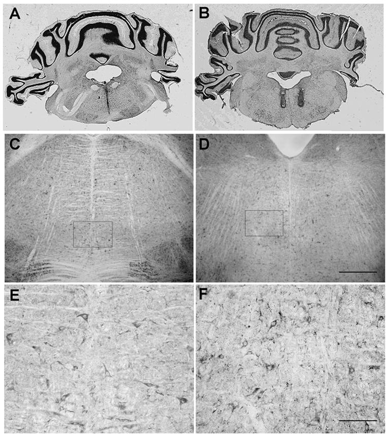
Photomicrographs showing placement of A) midline single cannula tract (bregma – 10.04 mm) and B) bilateral cannula tracts (bregma – 10.80 mm) within the RVM in brain sections counterstained with cresyl violet. Section thickness = 40 μm. Brightfield photomicrographs of Y1 receptor immunoreactivity in the RVM at C) bregma – 10.08 mm and D) bregma – 10.80 mm. Scale bars = 200 μm for C, D. The boxed areas in C and D represent areas of higher magnification as shown in E and F, respectively. Scale bars = 100 μm for E, F.