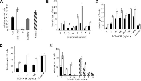
Figure 1.
Mutation of Tyr577 in βc of the GM-CSF/IL-3/IL-5 receptors enhances hematopoietic colony formation in response to GM-CSF. FL cells from βc−/−/βIL-3−/− mice were transduced with a retrovirus encoding the GM-CSF receptor α chain and either wild type βc or mutants of βc for 72 hours in a cocultivation system with ψ2 retroviral producer cells. Following transduction and quantification of transduction efficiency by immunofluorescence, 100 000 transduced cells were plated in 35-mm dishes containing 0.3% agar with human GM-CSF (100 ng/mL) or a cocktail of IL-6 (50 ng/mL), SCF (100 ng/mL), and Epo (4U/mL). (A) Comparison of wtβc with mutants in which all 8 tyrosines had been replaced by phenylalanine (F8), and the single-residue mutants Tyr577Phe or Ser585Gly in mediating colony formation in response to GM-CSF. This experiment is representative of 5 similar experiments. Error bars represent SEM from 3 to 5 replicate plates. ▨ depicts average colony formation of all groups in response to the cocktail stimulation. (B) TheTyr577Phe mutation (□) consistently enhanced colony formation from FL cells in response to 100 ng/mL GM-CSF compared with wtβc (■). Error bars represent SEM from 3 to 5 replicate plates. (C) Titration of human GM-CSF for its ability to stimulate colony formation in cells transduced with wtβc (■) or Tyr577Phe mutant (squlo]). Pooled data from 3 separate colony assays are shown. Error bars represent SEM from 3 to 11 plates per group. (D) Bone marrow (BM) cells from βc−/−/βIL-3−/− mice were transduced and plated in agar as above. Colony formation was stimulated with GM-CSF or the IL-6, SCF, and Epo cocktail as described in “Biological assays”. Colony formation was assessed at day 7, and in the experiment shown, error bars represent SEM from 6 replicate plates from 1 experiment that was representative of 3 performed. (E) FL cells transduced with either Tyr577Phe βc (□ and  ) or wt βc (■/▨) were cultured for the indicated times in liquid media of IMDM plus 15% HI-FCS supplemented with GM-CSF at 100 ng/mL (□ and ■) or a cytokine cocktail (50 ng/mL IL-6, 100 ng/mL SCF, and 10 ng/mL G-CSF;
  or ▨). At the indicated days, cells were counted, assessed for viability, and plated in agar for a further 10 to 14 days in cocktail containing SCF, Epo, and IL-6. Error bars represent SEM from 3 to 5 replicate plates.

