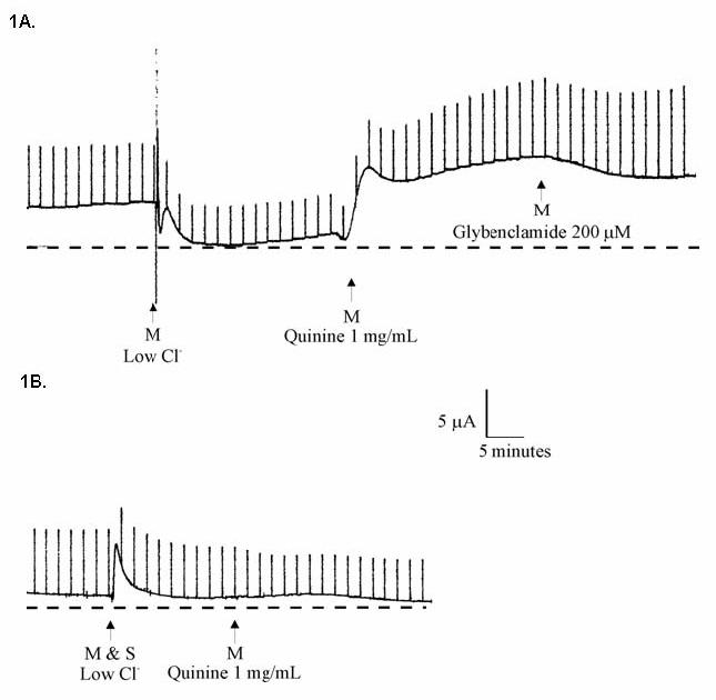
Figure 1. Apical addition of high dose quinine to Calu-3 monolayers.

Cells were grown at an air-liquid interface and studied in Ussing chambers. A: Calu-3 cells in Ringers solution followed by 1) mucosal (M) low- Cl− solution, 2) addition of 1 mg/mL quinine, and 3) addition of 200 μM glybenclamide. B: Same experiment as in A, with mucosal and serosal low- Cl− solution (i.e. no chloride gradient) prior to addition of quinine.
