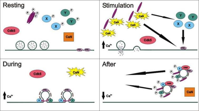
Fig 2. Model for the control of SVE by dynamin I phosphorylation.
(A) In a resting nerve terminal all phosphorylated dynamin I (purple bars) and its phosphorylation-dependent binding partner(s) (X & Y) are in the cytosol. Calcineurin (CaN) is inactive. (B) On nerve terminal depolarisation Ca2+ influx activates calcineurin, which dephosphorylates dynamin I and proteins X & Y. Dephosphorylation allows dynamin I to translocate to the plasma membrane via interactions with either or both phosphatidylinositol (4,5) bisphosphate (PIP2) or its phosphorylation-dependent binding partners (X & Y). (C) During depolarisation and SVE, dynamin I forms a collar round the neck of the retrieving SV and causes fission from the plasma membrane. (D) On termination of nerve terminal stimulation intracellular Ca2+ levels drop, inactivating CaN. This allows cdk5 to rephosphorylate dynamin I, X and Y. This rephosphorylation reduces its affinity for both PIP2 and proteins X & Y, facilitating the return of dynamin I to the cytosol for the next cycle of SVE.

