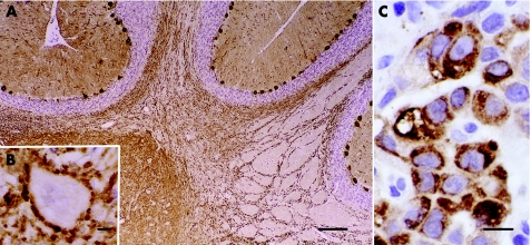
Figure 1 Frozen section of paraformaldehyde‐fixed rat cerebellum immunoreacted with the patient's serum. (A) Intense labelling of the cytoplasm, dendrites and axons of Purkinje cells. Bar = 120 μm. (B) Higher magnification showing a neurone of the deep cerebellar nucleus outlined by densely apposed axon terminals of Purkinje cells. Bar = 5 μm. (C) Paraffin‐wax‐embedded section of patient's non‐small‐cell lung cancer showing robust immunoreactivity when probed with a specific anti‐protein kinase Cγ antibody. Bar = 10 μm. Sections counterstained with haematoxylin.

An official website of the United States government
Here's how you know
Official websites use .gov
A
.gov website belongs to an official
government organization in the United States.
Secure .gov websites use HTTPS
A lock (
) or https:// means you've safely
connected to the .gov website. Share sensitive
information only on official, secure websites.
