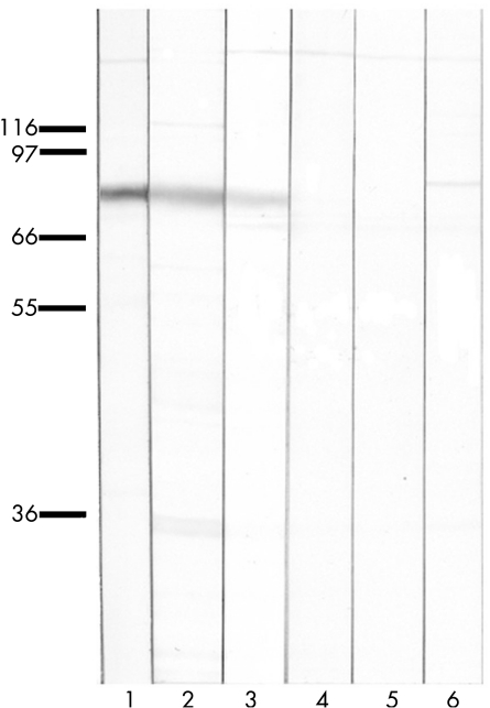
Figure 3 Immunoblots of rat cerebellum probed with an anti‐protein kinase Cγ (PKCγ)‐specific antibody (lane 1), patient's serum (lane 2), patient's immunoglobulin (Ig)G eluted from the PKCγ clone (lane 3) or an irrelevant clone (lane 4), IgG from an anti‐Hu‐positive serum eluted from the PKCγ clone (lane 5) and normal human serum (lane 6). Patient's serum and IgG eluted from the PKCγ clone recognise a band at 80 kDa also labelled with the anti‐PKCγ‐specific antibody. All lanes contain 10 µg of protein. All eluted IgGs normalised at 0.05 µg/ml.

An official website of the United States government
Here's how you know
Official websites use .gov
A
.gov website belongs to an official
government organization in the United States.
Secure .gov websites use HTTPS
A lock (
) or https:// means you've safely
connected to the .gov website. Share sensitive
information only on official, secure websites.
