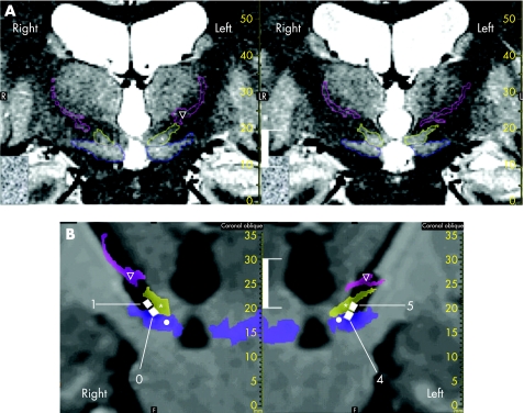
Figure 1 Effective contact locations (white vertical bar, 10 mm). (A) Determination of anatomical structures on stereotactic (stereotactic frame and location box in place) preoperative T2‐weighted slices (voxel size 0.6×0.5×2 mm3). Structures were identified according to their anatomical locations afforded by the image contrast, and surrounded manually on the coronal plane. Example of two joined coronal slices (right slice located 2 mm in front of the left slice); zona incerta (∇, light pink), subthalamic nucleus (*, yellow) and substantia nigra (•, blue). (B) Effective contact locations: white squares, contact numbers 0 and 1 for the right electrode and numbers 4 and 5 for the left electrode. Slices were reconstructed in pseudocoronal planes going through the electrode tracts. Anatomical structures identified on preoperative T2‐weighted slices were merged (voxel‐to‐voxel matching; semitransparent coloured surfaces) on postoperative T1‐weighted MRI (without stereotactic frame and location box; isotropic voxel size 1.3 mm3; electrodes can be seen in a black artefact form). Contacts are shown taking into account both the location of the centre, based on the postoperative radiograph stereotactic controls, and the zoom effect of the artefact (black signal; according to deep‐brain stimulation electrode geometry).

An official website of the United States government
Here's how you know
Official websites use .gov
A
.gov website belongs to an official
government organization in the United States.
Secure .gov websites use HTTPS
A lock (
) or https:// means you've safely
connected to the .gov website. Share sensitive
information only on official, secure websites.
