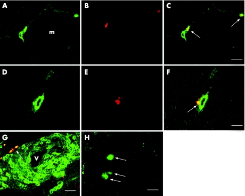
Figure 1 Granulysin expression in infiltrating cells in biopsy specimens of patients with polymyositis. (A–C) Granulysin is located in the cytoplasm of CD8 cells infiltrating around muscle fibres (denoted by m; arrows). (A) CD8 (fluorescein isothiocyanate (FITC)); (B) RC8 (Texas Red); (C) merged. Bar = 10 μm. (D–F) In this longitudinal section, granulysin (arrow) seems to have been released from a CD8 cell into the non‐necrotic muscle fibre located at the left. (D) CD8 (FITC); (E) RC8 (Texas Red); (F) merged. Bar = 5 μm. (G) Granulysin is occasionally expressed in CD4 cells surrounding blood vessels (denoted by v; arrow); CD4 (FITC)+RC8 (Texas Red). Bar = 15 μm. (H) Granulysin is also expressed in autoinvasive cells (arrows); RC8 (FITC). Bar = 10 μm.

An official website of the United States government
Here's how you know
Official websites use .gov
A
.gov website belongs to an official
government organization in the United States.
Secure .gov websites use HTTPS
A lock (
) or https:// means you've safely
connected to the .gov website. Share sensitive
information only on official, secure websites.
