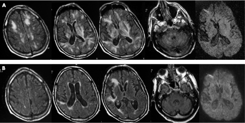
Figure 1 (A) Initial MRI findings. Fluid attenuated inversion recovery (FLAIR) sequence image showing diffuse white matter hyperintensity at both infra and supratentorial levels and in the basal ganglia. Hyperintensity extends from the periventricular white matter to the cortex. Diffusion‐weighted images (DWI)‐weighted MRI was normal. (B) MRI 3 weeks later. FLAIR sequence image showing dramatic improvement of the abnormalities but two areas of hyperintense signals in the right frontal region are shown without abnormalities on DWI‐weighted MRI.

An official website of the United States government
Here's how you know
Official websites use .gov
A
.gov website belongs to an official
government organization in the United States.
Secure .gov websites use HTTPS
A lock (
) or https:// means you've safely
connected to the .gov website. Share sensitive
information only on official, secure websites.
